
Understanding Respiratory Problems in Snakes
By Dr Michaela Betts
Respiratory problems are a commonly reported health concern in captive snake species. A study of 744 snake owners, surveyed worldwide in 2019, found 5.6% of participants reported signs of respiratory issues in at least one of their snakes. Suboptimal temperatures and humidity, recent movement, parasitism, malnutrition, and inappropriate ventilation are all identified stressors that can predispose to the development of respiratory disease. The following article will address some of the main causes and symptoms of respiratory problems in snakes, for expert and beginner keepers alike.

A boa under clinical assessment

Anatomy & Physiology
Snake species have a unique respiratory system compromising of nares, rhinarium, choana, larynx, trachea, bronchus/bronchi, and vascular and saccular lungs. They have no diaphragm and are unable to cough, so have no active way to remove airway mucus on a large scale. Their respiratory epithelium also has relatively few cilia compared to other species, so snakes also have a reduced ability to remove foreign material from the respiratory tract and clear mucous or inflammatory debris.
All snakes have a right lung which is divided into a cranial thick-walled vascular respiratory portion, and a caudal avascular thin-walled non-respiratory portion making an air sac or “saccular lung”. This non-respiratory portion is usually three to four times larger than the respiratory part and may act as an oxygen reservoir for periods of apnoea, or as a buoyancy aid in aquatic snake species. Boidae have a vestigial but functional left lung. Despite this, whilst respiratory disease is a common presentation in snakes, the larger, more sedentary Boids seem to experience issues more commonly than colubrid species.
Inspiration occurs through muscle groups expanding the ribs and lowering the intrapulmonary pressure, allowing a passive expiration as these muscles then relax. Whilst mammals are stimulated to breathe by high partial pressures of carbon dioxide, snakes are stimulated to breathe by low partial pressures of oxygen.
Clinical Signs
Respiratory infection may be seen as a sole issue, or in association with stomatitis (inflammation and/or infection of the oral mucous membranes). Reptiles can withstand prolonged periods of hypoxia (low levels of oxygen within the blood), and even anoxia (an absence of oxygen), and seem able to function in a somewhat normal physiological state even when in pulmonary compromise.
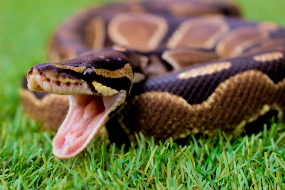
Clinical signs include audible wheezing or clicking noises when breathing, bubbles or discharge from the mouth or nares, open-mouth breathing, blue-tinged or pale gums, difficulty breathing, increased respiratory rate, yawning, and the snake resting in a more stretched-out position, or with their head and neck elevated. However, in some cases, especially Boids, the signs can be vaguer, and may just be lethargy and anorexia. There may also be a change in temperature preference noted, where the snake will suddenly start to favour the cooler areas of their set-up. This is because this will help to lower their metabolic rate and therefore enable them to better cope with the lower circulating oxygen levels.
Whilst it may be due to problems of the respiratory tract, it’s important to remember that difficulty breathing can also be seen in any disease process that is affecting a snake’s ability to expand its lungs, or that is compromising how well the lungs can function. For example, a space-occupying lesion within the coelom, such as an abscess or tumour, could impact lung expansion. Fluid build-up within the lungs, secondary to heart or liver disease, could also present as breathing difficulty. Therefore, whilst respiratory disease can be heavily suspected based on clinical signs, it is important to seek veterinary assistance from a snake-savvy vet to confirm the diagnosis and receive appropriate treatment.
A healthy royal python yawning.
Types of Respiratory Disease
Bacterial infection is a common cause of pneumonia in snakes and may be primary, or may be secondary to stomatitis, haematogenous spread, or infection of another organ in direct or close association, such as the liver. Infection may be focal, multifocal, diffuse, unilateral, or, in Boids, bilateral (focused on one spot, many spots, occurring down the side of the body or both sides of the body). Many types of pneumonia involve commensal bacteria of the respiratory tract, or environmental organisms that become pathogenic due to immunocompromise. Compared to healthy snakes, affected snakes seem to have heavy growths of aerobic gram-negative bacilli on culture and sensitivity such as Pseudomonas, Aeromonas, and Escherichia coli. However, some atypical bacterial infections can occur, such as mycobacteriosis, and these should be considered in persistent and recurring cases.
Fungal infection is a rare cause of pneumonia and is typically only seen in severely immunocompromised individuals or following prolonged systemic antibiosis. There are very few clinical cases reported in the literature, but multiple agents have been identified in both the skin and respiratory systems of snakes such as Aspergillus sp. and Candida sp.
Viral respiratory pathogens can be a primary cause of clinical and subclinical pneumonia in snakes, or appear as part of a mixed infection. A positive result does not necessarily mean they are a causative agent but could be contributing to overall immunosuppression. As such, viral infections should also be considered if recurrent or persistent outbreaks occur. Ophidian paramyxovirus has been a reported cause of pneumonia in many snake species, particularly in Viperids and Boids, with characteristic oedema and haemorrhage within the respiratory tract in some cases. The incurable arenavirus that causes Inclusion Body Disease in Boids has also been identified as a cause of secondary bacterial pneumonia. Other viruses to be aware of concerning respiratory disease are nidovirus in Boids, and reovirus and adenovirus, which are more commonly seen in Colubrids.
Parasitic infections affecting the respiratory system are reasonably common in snakes. Some parasites, such as Rhabdius, complete their lifecycle within the lungs. Others, such as Pentastomids, have larvae that migrate from ingested food into the lung, which then mature into adults there. These adults then reproduce within the lungs, and the larvae migrate up the trachea into the oral cavity where they are then swallowed and passed through the digestive system. There are also parasites, such as Ochetosomatidae, that inhabit the oral cavity and the adult parasites then migrate down the trachea to the lungs and air sacs. All parasites predispose their host to secondary bacterial pneumonia and migrating parasites can be associated with focal lesions within the lungs. Snake mites (Ophionyssus natricis), though ectoparasites, can also be a vector for some infectious causes of pneumonia, such as aeromoniasis.
Non-infectious causes of respiratory disease are uncommon but include trauma, particularly from penetrating injuries, neoplasia, and exposure to irritant gases or aerosols. Foreign bodies are incredibly rare, although inhalation of mucus or pus in cases of stomatitis, or occlusion of the airway from mucus plugs in dehydrated snakes, have been reported.
Predisposing Factors
Factors contributing to the development of respiratory disease in snakes include inappropriate humidity, inadequate temperature ranges, poor ventilation, stress and consequent immunosuppression, enclosure size, physiologic ecdysis, individual species characteristics, and habitat hygiene.
Inappropriately high or low humidity can affect ventilation and contribute to expediated bacterial and fungal growth in the environment, increasing the exposure for the snake and predisposing it to pneumonia. Equally, poor environmental hygiene can increase the bacterial and fungal exposure of a reptile over time, and lead to immunosuppression and opportunistic infection. Ectoparasites such as snake mites (Ophionyssus natricis) have also been linked to poor environmental hygiene.

Ophionyssus natricis
By Dack9 - Own work, CC BY 4.0, https://commons.wikimedia.org/w/index.php?curid=73207012
Preventative Measures
Prevention of respiratory disease involves good husbandry and quarantine measures, as well as prophylactic veterinary care.
Temperature & Humidity
Enclosure size is an important consideration for both thermoregulation and for enabling stretching out of the body. Many snakes develop clinical or subclinical bacterial pneumonias as a result of persistent suboptimal core body temperatures and consequent immunocompromise. Enclosures should be appropriately sized to provide the snake with the optimum temperature and humidity range for the species. As large as possible an appropriate temperature gradient is preferred to allow the snake to regulate its core temperature more effectively. Temperature and humidity should be monitored both where the heat is focused, and in the coolest region of the set-up as a minimum. A separate hygrometer should be used in each “microclimate” provided within the enclosure. Digital minimum-maximum thermometers are particularly useful as they will identify the highest and lowest temperatures reached in that area, not just display the current temperature.
Appropriate humidity is vital to thermoregulation and should not be overlooked, particularly in the larger tropical snake species. Less humid enclosures trap less heat and so are more at risk of significant fluctuations in temperature, particularly overnight. The viscosity of respiratory secretions is also increased in more dry environments, making them more likely to accumulate within the respiratory system if inappropriately dry. Conversely, inappropriately high humidity can impact the epithelial lining of the respiratory tract and make affected individuals more susceptible to developing a bacterial infection. Ventilation is key to helping avoid inappropriate rises in humidity and poor air quality.
Enclosure Sizing
Issues with thermoregulation can arise particularly in enclosures that are too small: heat sources can elevate the temperature throughout the set-up so that the snake is unable to cool down appropriately and more at risk of chronic dehydration. This dehydration is exacerbated by more rapid evaporation in warmer, smaller environments. There is also the potential for a relative increase in bacterial and/or fungal environmental contamination due to the smaller surface area of the set-up comparatively. Conversely, larger set-ups are more at risk of having inappropriately low temperatures. A combination of heat emitters and room temperature are usually employed for larger enclosures, and both air and surface temperatures within need to be monitored closely. Underheating should be avoided as it has been linked to reduced movement and therefore reduced clearing of discharges. It is also important to remember that heating underneath set-ups is not usually adequate for larger snakes. Heat burns are a real risk due to their weight and how snakes can remain on conductive heated surfaces for inappropriately long periods of time.
Set-ups should also, as a minimum, be large enough in design to allow a snake to stretch out its body completely. This reduces compression of the lungs and allows both the lung and air sac to fill and empty completely in a single respiratory cycle. Ideally, the set-up should also be large enough for appropriate exercise. Vertical space and the ability to climb are also important factors in arboreal and semiarboreal snake species, as gravity facilitates the drainage of exudative material through the glottis. This also better enables them to express normal behaviour.
Enclosure Cleaning
Appropriate hygiene and cleaning of set-ups is another important factor in preventing disease of any kind in captive snakes. Health issues most commonly arise in animals kept in enclosures that are difficult to clean, such as painted plywood, and where there is a high snake-to-cage size ratio so the snake is more likely to be in direct contact with its excrement. Frequent and thorough cleaning of the enclosure is necessary for all species, but the exact frequency required will depend on these factors. Substrates should always be replaced when soiled. Extra bowls and enrichment items are also recommended to allow rotation through the cleaning process with minimal stress to the animal. However, it is worth noting that porous items such as wood or cork may be impossible to appropriately disinfect, and it may be preferable to discard rather than rotate these items.
Few disinfectants are effective in the presence of organic matter such as excrement, so an appropriate cleaning regime should include the thorough cleaning of all surfaces and in-contact items with a mild detergent cleansing agent, such as warm soapy water, followed by a thorough rinse in hot water and then adequate drying, before application of an appropriate disinfectant. Disinfectants should be mixed and applied for the appropriate contact time according to the manufacturer’s instructions and should be selected for the type of enclosure being cleaned. Commonly used disinfectants include F10 disinfectant spray and 1:50 diluted bleach. The type of disinfectant used will also dictate if it should be rinsed off or not for optimum effect or to prevent toxicity. Good hand hygiene, such as using disposable gloves or alcohol-based hand rubs, to help prevent the spread of potential issues, should not be overlooked during the cleaning process.
Quarantine
Any new snake entering a collection should be quarantined upon arrival, regardless of source. This helps prevent the introduction of infectious agents into an established collection. Ideally, quarantined animals would be kept in a separate building and air-handling system from any other reptiles. However, where this is not possible, then a quarantine area should be as far away from the rest of the collection as possible. Quarantined animals should have separate tools and supplies that are not used for the rest of the collection. Their enclosure should be easy to disinfect with minimal substrate, ideally of easily disposable material like newspaper, but needs to meet the animal’s needs in terms of size, thermal gradient, humidity, and appropriate hides and climbing options. Porous items and surfaces should not be used in quarantine set-ups due to the difficulties of completely disinfecting them. Food items used on quarantine animals should be discarded if uneaten and not offered to any other animals, even those also in quarantine.
A 3-month minimum quarantine period, longer for wild-caught snakes or those with an unknown history (or known disease or parasitic infestation), is recommended to reduce the risk of a preventable outbreak within a collection. Animals should be weighed at regular intervals during this time, and their behaviour, demeanour, appetite, and overall condition noted. Ectoparasites should also be regularly checked for and treated accordingly if found. For snakes, some recommend they should eat for at least three feeds in a row before being considered well enough to leave quarantine, even if not displaying any clinical signs during their quarantine period.
At least two faecal samples throughout the quarantine should be screened for parasites. Viral screening with appropriate PCR and/or serological testing for the type of snake acquired is advised depending on the individual species’ susceptibility to certain viruses. It is recommended that new Boidae and Viperidae should be screened serologically for paramyxovirus, and Boidae also be screened for arenavirus, for instance. Depending on the known history of the snake, its source, and if they are displaying any clinical signs of disease, further investigations such as bloodwork or culture and sensitivity testing may be additionally indicated.
Summary
Respiratory disease in snakes is often multifactorial, with a large proportion of infections caused by commensal bacteria due to an individual being immunocompromised. Good husbandry, biosecurity, and quarantine measures are all contributing factors that help limit and prevent the spread and development of not only respiratory problems but other potential disease processes.

Jargon Buster
Epithelium – the thin tissue forming the outer layer of a body's surface and lining the alimentary canal (the whole passage of food from mouth to anus) and other hollow structures.
Cilia – Microscopic hair-like structures that vibrate to move fluid or provide propulsion.
Apnoaea – A temporary cessation of breathing (especially during sleep).
Boidae/Boids – Referring to the “boas” and “pythons” of the Boidae family.
Coelom – A fluid-filled body cavity that contains the organs, such as the lungs or heart.
Aerobic – Able to grow in the air.
Bacili – A type of bacterium.
Antibiosis – An antagonistic relationship between two organisms where one is harmed (the opposite of symbiosis).
Prophylactic – Intended to prevent disease.